In the study outlined below, it is clear that PRP inhibits bone matrix formation. this is consistent with previous reports. Perhaps, PRP is not useful in enchondral ossification but may be helpful in membranous ossification. Further research is needed.
APEX PRP.com
J Bone Joint Surg Am. 2007 Jan;89(1):139-47. Links
Platelet-rich plasma inhibits demineralized bone matrix-induced bone formation in nude mice.Ranly DM, Lohmann CH, Andreacchio D, Boyan BD, Schwartz Z.
It is unclear whether platelet-rich plasma is a clinically effective adjunct to osteoinductive agents such as demineralized bone matrix. It contains platelet-derived growth factor (PDGF), which decreases osteoinduction by human demineralized bone matrix in nude-mouse muscle, suggesting that platelet-rich plasma may also have a negative impact. This study tested the hypothesis that platelet-rich plasma reduces demineralized bone matrix-induced bone formation and that this effect varies with donor-dependent differences in platelet-rich plasma and demineralized bone matrix. Compared with platelet-poor plasma, platelet-rich plasma preparations exhibited a fourfold increase in the platelet count, a fifteenfold increase in the amount of transforming growth factor-beta, a sixfold increase in the amount of PDGF-BB, a fivefold increase in the amount of PDGF-AA, and a twofold increase in the amount of PDGF-AB. Platelet-rich plasma decreased the osteoinductivity of demineralized bone matrix implanted in immunocom-promised mice, and the activities of both demineralized bone matrix and platelet-rich plasma were donor-dependent. CLINICAL RELEVANCE: Platelet-rich plasma may not be an appropriate adjunct to demineralized bone matrix in some clinical applications.
Monday, January 15, 2007
Sunday, January 07, 2007
Platelet Rich Plasma and Meniscus Surgery
Here is the answer to another question about concerning PRP.
There is no data on the use of PRP for meniscus surgery so far. Many years ago, isolated meniscus repair was routinely enhanced using a fibrin clot which is similar to PRP. Again, this type of application makes sense but there isn't any solid information concerning its use in this area yet.
Adding PRP to meniscus repairs is best discussed on an individual basis with your surgeon.
Apex PRP.com
Platelet Rich Plasma and Degenerative Disc Disease
Recently, someone wrote in asking if PRP could help degenerative disc disease. Here is an answer to that question.
Presently, there are no specific human trials using PRP for degenerative disc disease. Theoretical and cell culture evidence does support its use. An article published in the esteemed journal Spine by Akeda et al stated:
"Platelet-rich plasma was effective in stimulating cell proliferation and extracellular matrix metabolism. The response to platelet-rich plasma was greater in the case of anulus fibrosus cells than of nucleus pulposus cells. The local administration of platelet-rich plasma might stimulate intervertebral disc repair. In addition, given the risks of using animal serum for tissue engineering, autologous blood may gain favor as a source of growth factors and serum supplements needed for stimulating cells to engineer intervertebral disc tissues."
Clearly, much more research is needed but the use of PRP for DDD does seem like a reasonable option that may one day be available.
Apex PRP
Saturday, January 06, 2007
Platelet Rich Plasma Enhances Nerve Repair
- In the study outlined below, Platelet Rich Plasma improved nerve repair. Specifically, "the data demonstrated a measurable neurotrophic effect when PRP was present, with the most favorable results seen with PRP added to suture".
This is yet another study that helps us understand how to best use PRP.
Allan Mishra APEX PRP
----------------------------------------------
- Laryngoscope. 2007 Jan;117(1):157-165.
Effect of Platelet Rich Plasma and Fibrin Sealant on Facial Nerve Regeneration in a Rat Model.
OBJECTIVE:: To investigate the effects of platelet rich plasma (PRP) and fibrin sealant (FS) on facial nerve regeneration. STUDY DESIGN:: Prospective, randomized, and controlled animal study. METHODS:: Experiments involved the transection and repair of facial nerve of 49 male adult rats. Seven groups were created dependant on the method of repair: suture; PRP (with/without suture); platelet poor plasma (PPP) (with/without suture); and FS (with/without suture) groups. Each method of repair was applied immediately after the nerve transection. The outcomes measured were: 1) observation of gross recovery of vibrissae movements within 8-week period after nerve transection and repair using a 5-point scale and comparing the left (test) side with the right (control) side; 2) comparisons of facial nerve motor action potentials (MAP) recorded before and 8 weeks after nerve transection and repair, including both the transected and control (untreated) nerves; 3) histologic evaluation of axons counts and the area of the axons. RESULTS:: Vibrissae movement observation: the inclusion of suturing resulted in overall improved outcomes. This was found for comparisons of the suture group with PRP group; PRP with/without suture groups; and PPP with/without suture groups (P < .05). The PRP without suture group had a significantly greater degree of recovery than the PPP without suture group (P < .05), but it did not have better performance than suture group (P > .05). The movement recovery of the suture group was significantly better than the FS group (P = .014). The recovery of function of the PRP groups was better than that of the FS groups, although this did not reach statistical significance (P = .09). Electrophysiologic testing: there was a significantly better performance of the suture group when compared with the PRP and PPP without suture groups in nerve conduction velocity (P < .05). The PRP with suture group had the best results when compared with the suture as well as the PPP with suture groups in duration and latency-2 of MAP (P < .05). For the FS groups, no results were found demonstrating a biological effect. The PRP with suture group demonstrated the best performance in the latency-2 and the area under the curve of MAP when compared with the suture and FS with suture groups (P < .05). Histomorphometric analysis: PRP with suture demonstrated the greatest increase in axon counts when compared with suture, FS with suture, and PPP with suture groups (P < .05). There was no statistically significant difference seen in axon diameter. CONCLUSION:: The best results for the return of function in our rat facial nerve axotomy models occurred when the nerve ends were sutured together. At the same time, the data demonstrated a measurable neurotrophic effect when PRP was present, with the most favorable results seen with PRP added to suture. There was an improved functional outcome with the use of PRP in comparison with FS or no bioactive agents (PPP). FS showed no benefit over conventional suturing in facial nerve regeneration. Our study provides the potential of a new clinical application for PRP in peripheral nerve regeneration.
Thursday, December 21, 2006
Stronger Tendons with Platelet Rich Plasma
This is an abstract of a recently published article about how PRP may produce stronger tendons. It represents just one of the many articles that are coming out about how PRP may actually work. Further research into this important topic is on-going in several centers.
Please visit Apex PRP for more information including video about Platelet Rich Plasma.
Acta Orthop. 2006 Oct;77(5):806-12.
How can one platelet injection after tendon injury lead to a stronger tendon after 4 weeks? Interplay between early regeneration and mechanical stimulation.
Virchenko O, Aspenberg P.
Orthopaedics and Sports Medicine, Department of Neuroscience and Locomotion, Linkoping University, Linkoping, SE-581 85, Sweden.
BACKGROUND: Mechanical stimulation improves the repair of ruptured tendons. Injection of a platelet concentrate (platelet-rich plasma, PRP) can also improve repair in several animal models. In a rat Achilles tendon transection model, 1 postoperative injection resulted in increased strength after 4 weeks. Considering the short half-lives of factors released by platelets, this very late effect calls for an explanation. METHODS: We studied the effects of platelets on Achilles tendon regenerates in rats 3, 5 and 14 days after transection. The tendons were either unloaded by Botulinum toxin A (Botox) injections into the calf muscles, or mechanically stimulated in activity cages. No Botox injections and ordinary cages, respectively, served as controls. Repair was evaluated by tensile testing. RESULTS: At 14 days, unloading (with Botox) abolished any effect of the platelets and reduced the mechanical properties of the repair tissue to less than half of normal. Thus, some mechanical stimulation is a prerequisite for the effect of platelets at 14 days. Without Botox, both activity and platelets increased repair independently of each other. However, at 3 and 5 days, platelets improved the mechanical properties in Botox-treated rats. INTERPRETATION: Platelets influence only the early phases of regeneration, but this allows mechanical stimulation to start driving neo-tendon development at an earlier time point, which kept it constantly ahead of the controls.
Wednesday, December 13, 2006
Platelet Rich Plasma Spine Disc Regeneration
PRP may also be useful for spinal disc regeneration.
"Platelet-rich plasma was effective in stimulating cell proliferation and extracellular matrix metabolism. The response to platelet-rich plasma was greater in the case of anulus fibrosus cells than of nucleuspulposus cells. The local administration of platelet-rich plasma might stimulate intervertebral disc repair. In addition, given the risks of using animal serum for tissue engineering, autologous blood may gain favor as a source of growth factors and serum supplements needed for stimulating cells to engineer intervertebral disc tissues." (Akeda et al Spine 2006)
For more information, please visit Apex PRP.com
Wednesday, November 29, 2006
Update on ABC TV Link
Please check out this link. We just check it and the ABC story is still available on it.
ABC TV Dean Edell Story on Platelet Rich Plasma
Watch this space for more updates on the research.
ABC TV Dean Edell Story on Platelet Rich Plasma
Watch this space for more updates on the research.
Saturday, November 11, 2006
ABC News TV spot on Platelet Rich Plasma
Recently, Dr. Allan Mishra's publication of the use of PRP for chronic elbow tendinosis was featured by Dr. Dean Edell's medical program on ABC news in San Francisco. To view this segment please visit Apex PRP.com and follow the link to the video.
It is a well produced piece that outlines the technique and some of the science behind the procedure.
It is a well produced piece that outlines the technique and some of the science behind the procedure.
Sunday, October 29, 2006
Forbes and Time Articles
Dr. Allan Mishra's article on the use of Platelet Rich Plasma for chronic elbow tendonitis (Tennis Elbow) has been outlined in Forbes and Time Articles.
For more information, please visit: Apex PRP.com
For more information, please visit: Apex PRP.com
Tuesday, October 24, 2006
News Articles on Tendonitis Paper
The publication of the paper using Platelet Rich Plasma has generated several articles. Here is a link to some of the stories via google: http://news.google.com/nwshp?hl=en&tab=wn&q=allan+mishra&ie=UTF-8&filter=0
More information can also be found at Apex PRP.com
More information can also be found at Apex PRP.com
Sunday, October 22, 2006
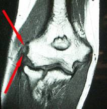
American Journal of Sports Medicine Publication
MRI of Chronic Elbow Tendinosis
Keep visiting this blog or Apex PRP.com for updates on Platelet Rich Plasma research and clinical applications.
Subscribe to:
Posts (Atom)